Building on our foundational work in the medial temporal lobe, the PATCH Lab has developed a comprehensive pipeline for integrating ultra-high resolution 7T postmortem MRI with whole hemisphere histopathology and enabling large-scale, spatially precise studies of neurodegeneration across the entire cerebral cortex.
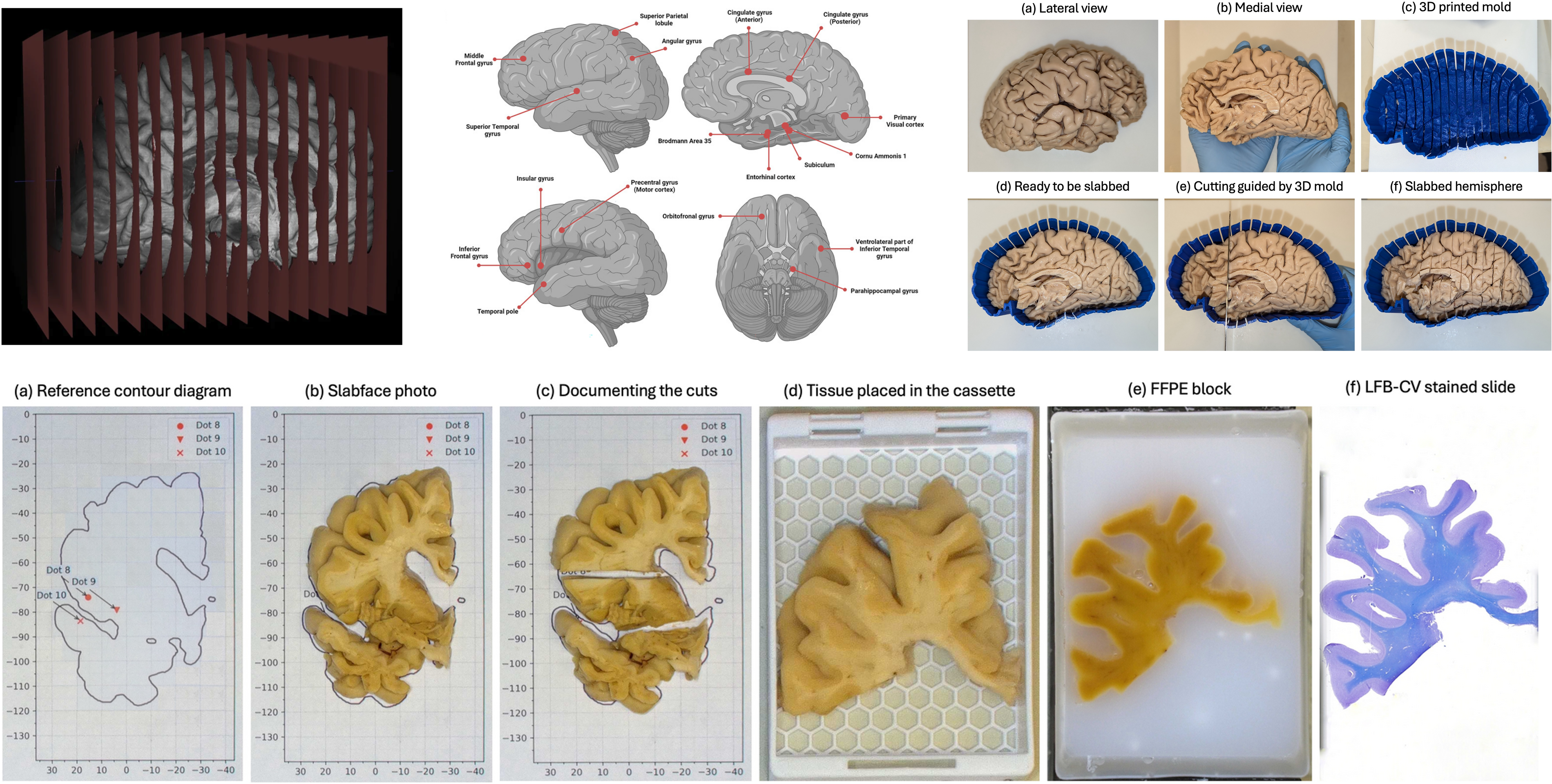
MRI-guided histology sampling protocol developed by PATCH Lab. We use patient-specific 3D-printed brain hemisphere cutting molds to guide our sectioning and staining strategy for each region of interest.
Traditional brain banking relies on sparse diagnostic sampling that can miss critical regions affected by Alzheimer’s disease and related disorders (ADRD). Our approach overcomes this limitation by using patient-specific 3D printed molds, generated directly from ex vivo MRI scans, to guide uniform 1 cm coronal slabbing of the hemisphere. This establishes a permanent spatial reference frame linking every histological section back to its precise location in the 3D MRI volume. We label 19 cortical landmarks on the MRI to target regions of known importance in ADRD, and process the corresponding tissue slabs for histological sectioning and staining at 20 micron thickness using both cytoarchitectural stains (LFB-CV, H&E) and immunohistochemistry (e.g., AT-8 for phosphorylated tau).
This pipeline is particularly well-suited for studying spatially heterogeneous, "patchy" pathologies such as cerebrovascular small vessel disease, chronic traumatic encephalopathy (CTE), aging-related tau astrogliopathy (ARTAG), and argyrophilic grain disease, at a precision and scale not previously feasible.
With over 250 hemispheres scanned to date and ongoing work to extend our quantitative pathology models to α-synuclein and TDP-43, this pipeline lays the groundwork for creating normative and disease phenotype maps of pathology, developing MRI-derived signatures applicable to in vivo biomarker development, and refining neuropathological sampling criteria across the full spectrum of neurodegenerative conditions (Athalye et al., 2025)
References
2025
-
Operationalizing postmortem pathology-MRI association studies in Alzheimer’s disease and related disorders with MRI-guided histology sampling
Chinmayee
Athalye, A.
Bahena, Pulkit
Khandelwal, Sheina
Emrani, W.
Trotman, Lisa
Levorse, Zahra
Khodakarami, Daniel T.
Ohm, Eric
Teunissen-Bermeo, N.
Capp, S.
Sadaghiani, Sanaz
Arezoumandan, Sydney A.
Lim, K.
Prabhakaran, R.
Ittyerah, John L.
Robinson, T.
Schuck, Edward B.
Lee, M.
Tisdall, Sandhitsu R.
Das, D.
Wolk, David J.
Irwin, and Paul
Yushkevich
Acta Neuropathologica Communications, May 2025
Cited by 8
Postmortem neuropathological examination, while the gold standard for diagnosing neurodegenerative diseases, often relies on limited regional sampling that may miss critical areas affected by Alzheimer’s disease and related disorders. Ultra-high resolution postmortem MRI can help identify regions that fall outside the diagnostic sampling criteria for additional histopathologic evaluation. However, there are no standardized guidelines for integrating histology and MRI in a traditional brain bank. We developed a comprehensive protocol for whole hemisphere postmortem 7T MRI-guided histopathological sampling with whole-slide digital imaging and histopathological analysis, providing a reliable pipeline for high-volume brain banking in heterogeneous brain tissue. Our method uses patient-specific 3D printed molds built from postmortem MRI, allowing standardized tissue processing with a permanent spatial reference frame. To facilitate pathology-MRI association studies, we created a semi-automated MRI to histology registration pipeline and developed a quantitative pathology scoring system using weakly supervised deep learning. We validated this protocol on a cohort of 29 brains with diagnosis on the AD spectrum that revealed correlations between cortical thickness and phosphorylated tau accumulation. This pipeline has broad applicability across neuropathological research and brain banking, facilitating large-scale studies that integrate histology with neuroimaging. The innovations presented here provide a scalable and reproducible approach to studying postmortem brain pathology, with implications for advancing diagnostic and therapeutic strategies for Alzheimer’s disease and related disorders.